A 30 YEAR OLD MAN WITH PAIN IN THE ABDOMEN ALONG WITH FEVER
THIS IS AN ONLINE E LOG BOOK TO DISCUSS OUR PATIENT'S DE - IDENTIFIED HEALTH DATA SHARED AFTER TAKING HIS / HER /GUARDIAN'S SIGNED INFORMED CONSENT .HERE WE DISCUSS OUR INDIVIDUAL PATIENT'S PROBLEMS THROUGH SERIES OF INPUTS FROM AVAILABLE GLOBAL ONLINE COMMUNITY OF EXPERTS WITH AN AIM TO SOLVE THOSE CLINICAL PROBLEMS WITH COLLECTIVE CURRENT BEST EVIDENCE BASED INPUT
A 30years old man driver by occupation came to the opd with chief complaints of fever since 6 days and pain abdomen since 3 days.
History of presenting illness:
Patient was apparently asymptomatic 6 days back then he developed High grade, intermittent fever along with chills and rigor which relieved temporararily medication.
2 days back he had dragging type of abdominal pain in right iliac ,right lumbar and right hypochondriac regions ,it was non radiating in nature
No h/o vomiting,loose stools,burning micturation,cough and shortness of breath.
PAST HISTORY:
NO H/O DM, HTN,CAD,Asthma,TB,Epilepsy
PERSONAL HISTORY:
DIET:Mixed
APPETITE:normal
BOWEL AND BLADDER movements: regular
SLEEP:adequate
ADDICTIONS: consumes toddy regularly and alcohol occasionally.
FAMILY HISTORY:
No significant family history
GENERAL EXAMINATION:
Patient is conscious,coherent,cooperative well oriented to time,place and person
He is moderately built and nourished
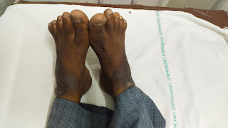
NO signs of pallor ,icterus,clubbing,cyanosis,lymphadenopathy,edema
VITALS at the time of admission:
TEMPERATURE:100°F
RR:17 cpm
PR:90 bpm
BP:130/80 mm Hg
SPO2:99 %
GRBS:185 mg %
SYSTEMIC EXAMINATION:
CVS: S1 and S2 are heard ,no murmurs
RESPIRATORY SYSTEM: BAE Present ,NVBS
P/A: Tenderness in right ilac and hypochondriac regions
CNS: NO focal neurological deficits .
INVESTIGATIONS:
HEMOGRAM:
Hb-13
TLC-13,700
Pl-2.6L
LFT:
Total Bilirubin-0.96
Direct Bilirubin-0.19
AST-38
ALT-39
ALP-134
TP-6.6
ALBUMIN-4.21
A/G-1.76
RFT:
Urea:-17
Creatinine:-1.0
Ca++-:9.6
Po4-:3.5
Na+:138
K+:4.5
Cl-:99
CUE:
ALB-nil
Pus cells:-2-3
Amylase-42.1
Lipase-21.4
RBS-116gm/dl
USG:
E/o 2.5x2.6cm heteroechoic lesion noted in left lobe of liver.
Liver abscess(with poor liquefaction-30%)
E/O 8-9mm hyperechoic focus noted in gall bladder— s/o gallbladder polyps.
E/O few freely mobile hyperechoic foci 1-2mm with posterior acoustic shadow noted in gall bladder— gallbladder microliths.


Comments
Post a Comment